
Pigeon Fever
What is pigeon fever?
Takeaways
- The causative bacteria, Corynebacterium pseudotuberculosis, can live in the ground, feces, hay and shavings for long periods.
- Horses are infected when bacteria gain access to the body through small scrapes or wounds in the skin, either by direct contact or by insects.
- Pigeon fever occurs in 3 main forms: external abscesses, internal abscesses, and ulcerative lymphangitis.
- Fly control is key to prevention and control of pigeon fever.
- Infected horses should be isolated when possible and care should be taken to avoid transmission of the bacteria to healthy horses on hands, clothing, and equipment such as buckets and grooming tools.
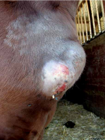
Pigeon fever, also called “dryland distemper” or “false strangles”, is an infection caused by the bacteria Corynebacterium pseudotuberculosis that typically causes large abscesses to form on the chest region of the horse or under the belly. The swelling on the horse’s chest resembles a pigeon’s breast, which is how the disease got its name. The bacteria lives in the ground, where it can survive for long periods. It can also survive for shorter periods in hay and shavings. Horses are infected when bacteria gain access to the body through small scrapes or wounds in the skin, either by direct contact with contaminated soil or objects, or by insects (flies) that deposit the bacteria on broken skin. Pigeon fever can occur year-round, with seasonal peaks of cases occurring during the summer through early winter, which could be indicative of high insect activity and the lengthy incubation period (1 - 4 weeks) for infected horses to develop clinical disease.
What are the clinical signs of pigeon fever?
Pigeon fever occurs in three main forms:
- External abscesses – frequently in the chest region or along the abdomen, but may also occur on the mammary gland, groin area, prepuce, triceps, limbs, and head
- Internal abscesses – most commonly seen in the liver, spleen, kidneys, and lungs
- Ulcerative lymphangitis (limb infection along the lymphatic system) – a painful infection of the lymphatics, most often involving the hind limbs and causing swelling and oozing sores
Depending on the form of the disease, an infected horse may also exhibit fever, lameness, weight loss, decreased appetite, lethargy, signs of respiratory disease, and abdominal pain. On rare occasions, the bacterium can cause osteomyelitis (infection of the bone) or septic arthritis (an intensely painful infection in a joint that carries a poor prognosis unless detected early and treated aggressively).
How is pigeon fever diagnosed?
Diagnosis is based on clinical signs, along with time of year, prevalence of the disease in the geographic location, and historical information. Definitive diagnosis is often made by culturing drainage from an abscess to determine if the Corynebacterium pseudotuberculosis bacteria is present. Ultrasound can be used to guide sampling of deep abscesses. It is important to confirm an accurate diagnosis based on bacterial culture to initiate the appropriate treatment. Not all abscesses are caused by Corynebacterium pseudotuberculosis.
Diagnosis of internal abscesses can be difficult and is based on clinical signs, laboratory testing including an antibody test (i.e. synergistic hemolysin inhibition, or SHI test, which looks for an immunological response to the bacterial exotoxin in the serum of the patient), diagnostic imaging (ultrasound), and bacterial culture.
For horses with ulcerative lymphangitis, bloodwork and antibody titers (i.e. SHI titers) are obtained and skin biopsies from ulcerative lesions are cultured for Corynebacterium pseudotuberculosis.
How is pigeon fever treated?
The severity of external abscesses can vary widely, but most straightforward cases are treated with hot compresses, poultices, lancing and draining, with collection of the infected material. This is preferable in order to prevent the discharge from contaminating the environment as the abscess slowly drains. Nonsteroidal anti-inflammatory drugs (NSAIDs) can be administered to ease discomfort before or after the abscess is drained. Timely intervention is key to the resolution of clinical signs and to prevent secondary complications. Antibiotics are not necessary to treat external abscesses in most horses and may prolong time to resolution. Antibiotic therapy may be justified with prolonged infection, and these cases warrant further discussion with your veterinarian.
Treatment for horses with internal abscesses includes long-term antibiotic therapy.
Horses with ulcerative lymphangitis or cellulitis should be treated early and aggressively with antibiotics to prevent residual lameness or limb swelling. Typically, intravenous antibiotics alone or in combination with an oral antimicrobial are used until lameness and swelling improve. Subsequently, orally administered antibiotics are continued to prevent relapse. Physical therapies, such as hydrotherapy, hand walking, and leg wraps, are often recommended.
What is the prognosis for pigeon fever?
The prognosis for horses with external abscesses is very good; most patients recover within 2 - 3 weeks. With internal infections, the prognosis can range from guarded to good. Early recognition and appropriate long-term (2-3 months, sometimes more) antibiotic therapy offer the best chance for complete recovery. Infections within the abdominal cavity are fatal if left untreated. Horses with ulcerative lymphangitis can have some residual lymphatic damage if not treated early and aggressively, which can make them prone to limb swelling and recurrence of infection. Most cases resolve within a month, but chronic swelling of the limbs and lameness can persist.
How can pigeon fever be prevented?
Fly control, using feed-through products and/or fly repellents, especially on horses with open wounds or draining abscesses, is key to prevention and control of pigeon fever. As flies can cause skin inflammation on the chest or in the girth area of some sensitive horses (i.e. midline dermatitis), fly control using fly sheets, spray or ointments is important. Regular manure management and sanitation programs should also be established in order to control insect populations.
Quarantine and observe new horses for signs of infection before they are introduced into the resident population. Isolate known infected horses when possible and practical, wash/sanitize hands after handling infected horses, and use gloves and protective outerwear. Avoid using the same items (buckets, pitchforks, and other materials) for infected horses and the general horse population. Carefully clean and disinfect areas potentially contaminated by pus from draining abscesses. Inspect stalls, paddocks and fields for sharp edges or objects that could cause wounds on your horse’s skin, which might subsequently become infected.
No further precautions are needed once infected horses are recovered and there is no drainage from abscesses. The bacterium is unfortunately endemic in many areas in California as there is no practical way to eliminate the bacteria from soil. There is currently no vaccine for pigeon fever. Horses that recover from the disease are often less likely to be affected again the future.
For more information:
UC Davis Center for Equine Health Horse Report, Winter 2014
California Animal Health and Food Safety Laboratory: Testing
*This article may not be reproduced without the written consent of the UC Davis Center for Equine Health. Please email requests to [email protected].